UNIT 1: CHEMEIA OF THE BIOS
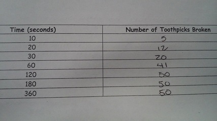
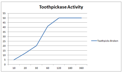
G-TOOTHPICKASE ACTIVITY: 08-13-14

Questions and Answers:
1. a. In this model, what is the substrate? The substrate is the toothpicks.
b. What acts as the enzyme? The person is the enzyme.
c. What is the product? The product is the broken toothpick.
2.During what time period was your reaction rate the fastest? Remember reaction rate can be found from the slope of the graph or number of toothpicks broken per time period.
The fastest time period was the 30 second time period that is when the amount raised the most.
3. What happens to the reaction rate as the supple of toothpicks runs out?
The reaction rate stayed the same.
4. What would happen to the reaction rate if the toothpicks were spread out so that the "enzyme" has to reach for them (substrate concentration is decreased)?
The reaction rate would slow down because the "enzyme" would have to reach to find the toothpicks.
5. What would happen to the reaction rate if more toothpicks were added?
The reaction rate would go down because the "enzyme" would get tired more easily.
6. How could we increase enzyme concentration? What would happen to the reaction rate?
You could increase the enzyme concentration by adding more people to help break the toothpicks. The reaction rate would increase because of the extra help.
7. How could we represent a competitive and noncompetitive inhibitor? How would these affect reaction rates?
The competitive inhibitor would be trying to break the toothpicks as fast as they could, while the noncompetitive inhibitor would be going at their own pace. The reaction rate of the competitive inhibitor would be much higher than that of the noncompetitive inhibator.
|
|
G-LAB 8: PINEAPPLE ENZYMES & JELLO MOLDS: 08-15-14
PROCEDURE:
1. gather all supplies:
-3 beakers
-10 test tubes (5 per trial)
-hot plate
-frozen, fresh, canned and cooked pineapple
- jello
-stirring rods and knives
2. Follow the instructions on the jello box to make jello.
3. pour jello (liquid) into the test tubes (one inch)
4. Put one piece of each kind of pineapple in each kind of test tube (leave last test tube with out pineapple)
5. Let sit until jello forms
6. retrieve jello and record results
7. repeat steps 2-6 for second trial.
Hypothesis: If we drop the pineapple in the jello, we believe the enzymes will break down the jello and not let it set.
Independent Variable: Jello
Measurement of Independent Variable: About an inch in each test tube.
Number of Trials: 2
Dependent Variable: Pineapple
Measurement of Dependent Variable: About 5 grams of Pineapple
Control: Non pineapple jello
Other Controlled Factors: Same fridge, same jello mix, same temperature, same amount of time in the fridge, and same size pineapple
Questions and Answers:
1. Clearly describe the result of your experiment. In which test tubes did the gelatin jell. which did not?
The results of the experiment:
Fresh Pineapple- The jello didn't set
Canned Pineapple- The jello set fully
Frozen Pineapple- The pineapple dissolved in the jello and the jello didn't set
Cooked Pineapple- The jello set fully
2. Clearly explain the result of your experiment. Why did some test tubes of gelatin jell, why did others not. Be specific!
The fresh and frozen pineapple have the enzyme bromelain which breaks the jello down. The canned pineapple set because of the preservatives that is was sitting in. The cooked set because the enzyme was cooked out of the pineapple.
3. What is the enzyme in your experiment?
The enzyme is the bromelain that is in the pineapple.
4. What is the substrate in your experiment?
The substrate is the test tube because that is where the reaction is taking place.
5. What is (are) the product(s) in your experiment?
The product is the solid or liquid jello.
6. What type of organic molecule is gelatin?
Gelatin is a carbohydrate because of the short term energy it gives.
7. What type of organic molecule is bromelain?
Bromelain is a protein because it speeds up reactions and it isn't consumed.
8. Write a "word equation" to describe the chemical reaction that occurs when pineapple is mixed with gelatin.
Fresh + Jello + Enzyme = Unset Jello
Frozen + Jello + Enzyme = Unset Jello
Canned + Jello - Enzyme = Set Jello
Cooked + Jello - Enzyme = Set Jello
9. Is the reaction of bromelain and gelatin dehydration synthesis or hydrolysis? Explain.
The reaction is dehydration synthesis because the enzyme breaks down the bonds of the water molecules.
10. Why were the results of the freshly cooked pineapple different that the results of the fresh, raw pineapple? Be specific!
Cooking the pineapple heated the enzymes up and destroyed them. The enzyme stayed untouched in the fresh pineapple.
11. What is meat tenderizer and what does it do?
Meat tenderizer has bromelain and it breaks down and digests protein.
12. Pineapple Enzyme denaturing experiment:
Procedure:
1. Supplies:
20 test tubes
fresh pineapple
a timer
1 knife (cutting the pineapple)
jello
a scale
Hydrogen Peroxide
2. Cut 8 pieces of pineapple into equal size and weight pieces.
3. Place 1 piece of pineapple in each test tube.
4. Place sets of 1 test tube of pineapple in 10 different temperature places. (10*F, 20*F, 30*F, 40*F, 50*F, 60*F, 70*F, 80*F, 90*F, 100*F)
3. Set the timer for 24 hours.
4. Take out the pineapple and lay them out in order of temperature.
5. For each test tube, add 10 ml of hydrogen peroxide to each test tube and record your results on a data table on a scale of 0-10 (10 being highly reactive and 0 being no reaction.)
6. Take 10 new pineapple slices and place it in the 10 other test tubes.
7. For the first temperature you got a 0 reaction on, take the previous 9 temperatures to use for the new data. (ex: if the temp with a 0 reaction rate was 70*F, you would take the test tubes and put them at the new temperatures of 60*F-70*F)
8. Repeat steps 3, 4, and 5.
9. The first temperature with 0 reaction rate if the temperature the pineapple enzyme will denature.
Fresh Pineapple, Canned Pineapple, Frozen Pineapple, Cooked Pineapple, Normal Jello
G-ENZYME LAB: 08-20-14
Part A:
1. The gas being released by the liver in the hydrogen peroxide is oxygen.
2. The test tube has gotten colder, this determines that the reaction is exothermic.
3. The liquid in the test tube that we poured off is water.
4. If we added more liver to this solution no reaction would happen.
5. When we put more liver into the poured off solution the reaction tested as a 0.
6. When we put more Hydrogen Peroxide on the liver used in the first test tube the reaction rate was a 0.
Part B:
Rate of reaction (0-5)
Potato
0 0
Apple
0 0
Chicken
1 1
7. The tissue the contains catalase is the chicken liver.
8. Yes some contain more catalase than others because the reaction is greater.
Rate of reaction (0-5)
Potato
0 0
Apple
0 0
Chicken
1 1
7. The tissue the contains catalase is the chicken liver.
8. Yes some contain more catalase than others because the reaction is greater.
Part C:
9. The boiling of the liver will make the enzyme denature.
10. The reaction rate of the boiled liver and peroxide is 0.
11. The reaction rate of the cold liver and cold peroxide is 0.
12. The reaction rate of the warm liver and warm peroxide is 0.
9. The boiling of the liver will make the enzyme denature.
10. The reaction rate of the boiled liver and peroxide is 0.
11. The reaction rate of the cold liver and cold peroxide is 0.
12. The reaction rate of the warm liver and warm peroxide is 0.
Part D:
13. The reaction rate of test tube 1 was 4.
14. The reaction rate of test tube 2 was 2.
15. The reaction rate of test tube 3 was 0.
16. The reaction rate of test tube 4 was 0.
17. The reaction rate of test tube 5 was 0.
18. The optimal pH for catalase is between 1 and 6.
13. The reaction rate of test tube 1 was 4.
14. The reaction rate of test tube 2 was 2.
15. The reaction rate of test tube 3 was 0.
16. The reaction rate of test tube 4 was 0.
17. The reaction rate of test tube 5 was 0.
18. The optimal pH for catalase is between 1 and 6.
Part E:
For the new experiment, we would take different materials such as different fruits and meats. Also 2 to 3 different chemicals ( HCl, NaOH). Then carefully put each of the fruit or meats into a test tube of each of the new chemicals. Then, add more of each chemical after each reaction has stopped, and record the results.
For the new experiment, we would take different materials such as different fruits and meats. Also 2 to 3 different chemicals ( HCl, NaOH). Then carefully put each of the fruit or meats into a test tube of each of the new chemicals. Then, add more of each chemical after each reaction has stopped, and record the results.
Data Analysis:
1. Describe the relationship between catalase and hydrogen peroxide. Indicate which is the enzyme, which is the substrate and what occurs during the reaction. (2)
Catalase breaks down hydrogen peroxide. Catalase is the enzyme, hydrogen peroxide is the substrate. The liver molecules were broken down.
2. Is catalase reusable? Use your data to support your answer. (1)
No, catalase is not reusable. When we put liver that had already been used in a reaction into new peroxide, no reaction occurred.
3. How does temperature and pH affect the reaction of catalase? Propose a way to refine your experiment to find the exact, or OPTIMAL pH and temperature of catalase. (3)
Temperature and pH affects how fast or slow the reaction happens. To find pH, you should use different chemicals and use a thermometer to find the temperature.
4. Amylase is an enzyme that can be used to break down carbohydrates, like those found in bread and crackers to individual units of sugars. This is why bread begins to taste sweet as we chew it, amylase is found in saliva. Benedict's solution is a chemical that changes color in the presence of sugar. How would you design an experiment to test the reaction rate of amylase in breaking down starch into sugars? (2)
For the experiment: first I'd have to obtain several different types of sugar and Benedict solution. Also I would need multiple test tubes to do multiple trials in. I would put each of the different sugars into test tubes, then add the Benedict's solution. Then add the amylase and set a timer to see how long it takes. I'd also want a control of sugar and Benedict solution to compare the results against.
S-PATTERN MATCHING: 08-24-14
I took the molecules and separated them by structure. Then I looked at the atoms that make them up and broke them into groups based on the main atom. Then I broke those down by the atoms that bonded to the main atom.
B-PRION READING: 08-24-14
Prions are infectious protein that cause many nuerodegenerative diseases. To study prions scientists use synthetic model but the models are not as complete as the real thing. Prion are three sided and cylindrical, they do not have DNA or RNA so the way they reproduce is by making other proteins fold incorrectly.
S-COACERVATES READING: 08-25-14
This reading was about how RNA reacts in aqueous two-phase solutions and coacervates droplets. The article talked about how the results were found, how the group did the lab to find the results. Then they went on to explain why they got the results they did. How the RNA went across the droplet boundary with each of the chemicals used. The grouped talked about the chemicals used and what they are. They finished with references to help people better understand what they experimented.
G-ACTIVITY 1: DETERMINE THE REACTION RATE IN THE PRESENCE OR ABSENCE OF AN ENZYME
Analysis:
Since we did not get a reaction we feel we did something wrong. Some of the things that could have went wrong were not making the solutions strong enough or using the wrong amounts of the buffer, enzyme, or substances. A couple other things could have been inconsistent time or using expired chemicals.
Since we did not get a reaction we feel we did something wrong. Some of the things that could have went wrong were not making the solutions strong enough or using the wrong amounts of the buffer, enzyme, or substances. A couple other things could have been inconsistent time or using expired chemicals.
G-MACRO-MOLECULES LAB: 09-02-14
Analysis Questions:
1. Was the data collected in this lab qualitative or quantitative? Explain
Qualitative because their was physical color changes.
2. What are some limitations in using the indicators to test for the various carbon-based molecules? Include at least 2 limitations.
One limitation is that only one color was used to indicate a reaction happened and another is that the solution could only get as dark as the indicator used.
3. If we did this lab again, how could you minimize errors? How could you get a more accurate idea of what carbon - based molecules are in various substances?
To minimize errors we could make sure the temperature is perfect for carbon-based molecules. To get a more accurate idea we could use more carbon-based solutions to see how they react.
Conclusion:
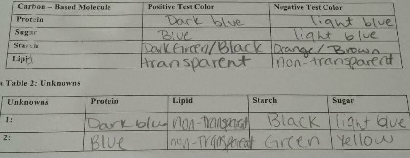
Claim: The unknown solutions are starch and sugar.
Evidence: They evidence is that the unknown solutions reacted the most with the indicator of that organic material.
Reasoning:
1. Was the data collected in this lab qualitative or quantitative? Explain
Qualitative because their was physical color changes.
2. What are some limitations in using the indicators to test for the various carbon-based molecules? Include at least 2 limitations.
One limitation is that only one color was used to indicate a reaction happened and another is that the solution could only get as dark as the indicator used.
3. If we did this lab again, how could you minimize errors? How could you get a more accurate idea of what carbon - based molecules are in various substances?
To minimize errors we could make sure the temperature is perfect for carbon-based molecules. To get a more accurate idea we could use more carbon-based solutions to see how they react.
Conclusion:
Claim: The unknown solutions are starch and sugar.
Evidence: They evidence is that the unknown solutions reacted the most with the indicator of that organic material.
Reasoning:
UNIT 2: KITARO
G-LAB 4: MEETING THE "PROTISTS": 09-12-14
Part 1: The Chromalveolates
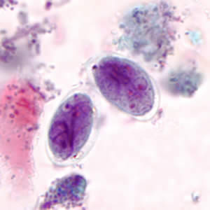
1A: The Apicomplexans
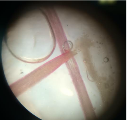
Slide: Plasmodium spp.
Identify: life cycle stage that you are looking at under the microscope.
Slide: Plasmodium spp.
Identify: life cycle stage that you are looking at under the microscope.
The life cycle stage is the Trophozoite Stage.
1B: The Ciliates
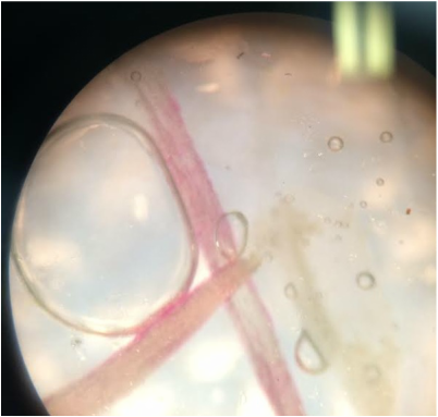
Slide: Paramecium Caudatum, Vorticella
Identify:
Macronucleus is the larger of the two nuclei in Ciliates.
Oral Groove is the site where food is taken in to the cell.
Cilia is a haor-like structure that moves the cell and debris.
Food Vacuole is a membrane enclosed organelle that digests food for the cell.
Contractile Vacuole pumps fluids from inside the cell to the outside and keeps the cell at its equilibrium.
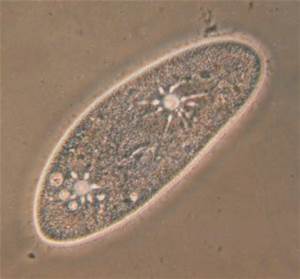
Paramecium Caudatum
|
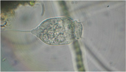
Vorticella
|
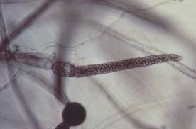
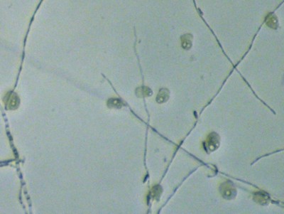
1C: The Oomycetes (Water Molds or Egg Molds)
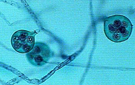
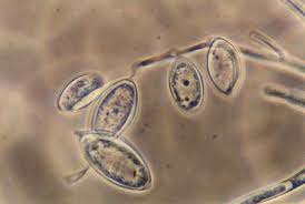
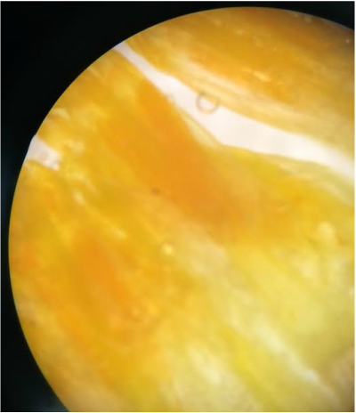
Slide: Saprolegnia
Identify:
Zoosporangia is a sporangium (spore case) in which zoospores are produced.
Oogonium is one of the undifferentiated germ cells giving rise to oocytes or the one-celled female reproductive organ in certain thallophytes, usually a more or less spherical sac containing one or more eggs.
Antheridium is a male reproductive structure producing gametes, occurring in ferns, mosses, fungi, and algae.
Part 2: The Plantae
2A: The Chlorophytes (Green Algae)
Find: Chlamydonmonas spp., Spirogyra spp., Volvox spp., Ulva spp.
Find: Chlamydonmonas spp., Spirogyra spp., Volvox spp., Ulva spp.
Part 3: The Excavates
3A: The Diplomonads
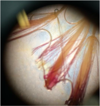
Find: Giardia Lamblia
Find: Giardia Lamblia
Giardia Lamblia
3B: The Kinetoplastids
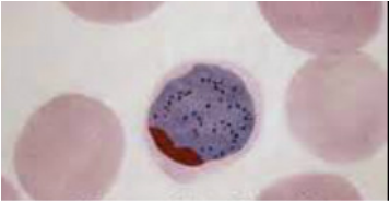
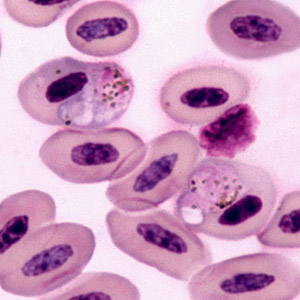
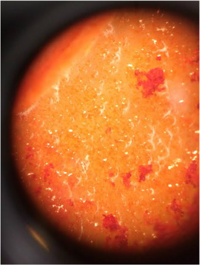
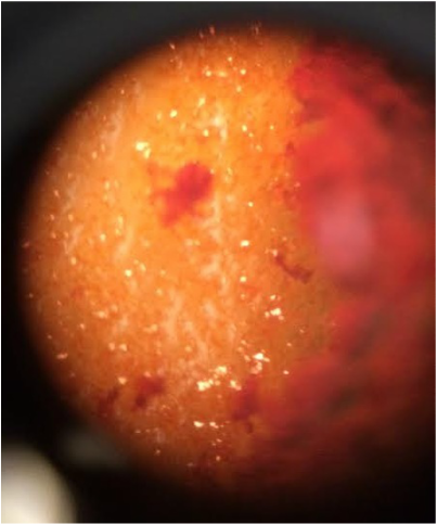
Slide: Trypanosoma (Note the Trypanosoma are in a blood smear)
Identify:
Nucleus is a specialized mass of protoplasm encased in a double membrane and found in most living eukaryotic cells (directs their growth, metabolism, reproduction, and functioning in the transmission of genic characters).
Flagella is a long, lashlike appendage serving as an organ of locomotion in cells.
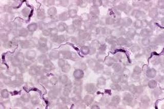
Trypanosoma
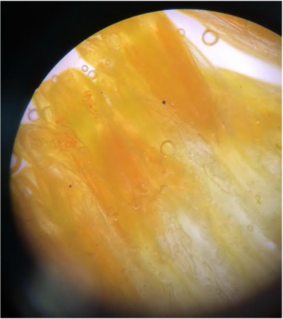
Part 4: Rhizaria
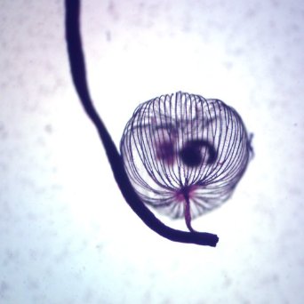
4A: The Foraminiferans
Find: Foraminifera Strewn
Find: Foraminifera Strewn
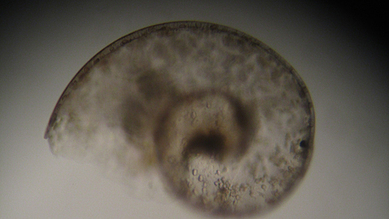
Foraminifera Strewn
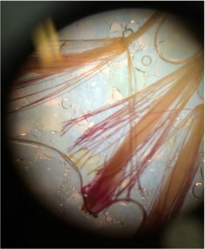
4B: The Radiolarians
Find: Radiolaria Strew
Find: Radiolaria Strew
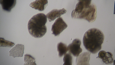
Radiolaria Strew
Part 5: Unikonts
5A: The Loboseans
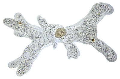
Slide: Amoeba Proteus
Find Amoeba spp.
Slide: Amoeba Proteus
Find Amoeba spp.
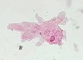
Amoeba
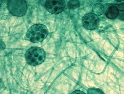
5B: The Plasmodial Slime Molds
Slide:Dictydium spp.
Identify:
Plasmodium is an ameboid mass (sheet) of cytoplasm characterized as some stage of an organisms or any parasitic protozoan of the genus Plasmodium (can cause malaria in humans).
Sporangia is the case (sac) in which spores are produced.
Slide:Dictydium spp.
Identify:
Plasmodium is an ameboid mass (sheet) of cytoplasm characterized as some stage of an organisms or any parasitic protozoan of the genus Plasmodium (can cause malaria in humans).
Sporangia is the case (sac) in which spores are produced.
5C: The Cellular Slime Molds
Identify:
Amoebas are any one-celled protozoa of the order Amoebida with a nucleus surrounded by cytoplasm that forms temporary extensions by which the organism moves, engulfs food particles, and forms food vacuoles.
Pseudoplasmodium is an amoeboid with many diploid nuclei that are the results of many nuclear divisions without cytokinesis and normally referred to as the feeding stage of macroscopic slime molds.
Fruiting Body (Sorocarp) is an organ that produces spores (fructification).
G-RED ONION OSMOSIS LAB: 09-15-14
Conclusion Questions:
1. Draw a diagram to indicate the relative proportions of salt and water within the onion cells and outside the onion cells when they were placed in the saline (salt) solution. Also use an arrow to properly indicate the direction of osmosis.
1. Draw a diagram to indicate the relative proportions of salt and water within the onion cells and outside the onion cells when they were placed in the saline (salt) solution. Also use an arrow to properly indicate the direction of osmosis.
Cell in a saline (salt) solution
75% water out, 25% salt in
2. Draw a diagram to indicate the relative proportions of salt and water within the onion cells and outside the onion cells when they were placed in the distilled water solution. Also use an arrow to properly indicate the direction of osmosis.
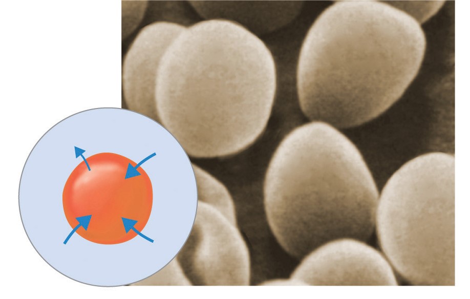
Cell in a distilled water solution
25% salt out, 75% water in
Application Questions:
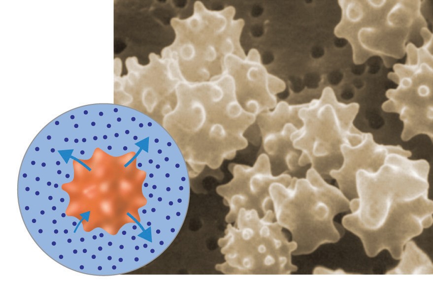
3. Red blood cells (and other animal cells) placed in a distilled water solution usually swell up and burst. Why prevented the red onion cells from swelling up and bursting when they were placed in the distilled water?
The red onion cells didn't swell up and burst because it is a plant cell, not an animal cell, where plants need to be submerged in water to stay alive they filter excess water out.
The red onion cells didn't swell up and burst because it is a plant cell, not an animal cell, where plants need to be submerged in water to stay alive they filter excess water out.
4. Why do grocery store owners spray fresh fruits and vegetables with water?
It keeps the fruits and vegetables hydrated so they don't shrivel and die.
It keeps the fruits and vegetables hydrated so they don't shrivel and die.
5. Roads are sometimes salted to melt ice. What does this do to plants around the roadside and why?
The salt kills the plants, the salt steals the water out of the cell.
The salt kills the plants, the salt steals the water out of the cell.
6. If a shipwrecked crew drinks salt water, they will probably die. Why?
The salt water will dehydrate the cells, making them shrivel and lose the ability to function properly.
The salt water will dehydrate the cells, making them shrivel and lose the ability to function properly.
7. If a bowl of fresh strawberries is sprinkled with sugar, a few minutes later the berries will be covered with juice. Why?
The sugar is acting the same as salt does on a cell, it draws out the liquid to the surface.
The sugar is acting the same as salt does on a cell, it draws out the liquid to the surface.
G-ALCOHOLIC FERMENTATION IN YEAST: 09-19-14
1. Use the terms carbon dioxide and oxygen to complete the following equation to describe aerobic respiration.
Glucose + Oxygen -> Carbon dioxide + water + oxygen
Glucose + Oxygen -> Carbon dioxide + water + oxygen
Experiment 1 - Effects of Sucrose Concentration on the Rate of Alcoholic Fermentation in Yeast
1. Humans use yeast everyday to make bread, wine, and beer. What is yeast?
Yeast is a fungus that turns sugar into alcohol.
Yeast is a fungus that turns sugar into alcohol.
2. What is sucrose?
Sucrose is a sugar or carbohydrates.
Sucrose is a sugar or carbohydrates.
3. In your experiment, you will grow yeast in a test tube filled with water and sealed with a balloon. Do you think these growth conditions are aerobic or anaerobic?
I think the growth conditions are anaerobic.
I think the growth conditions are anaerobic.
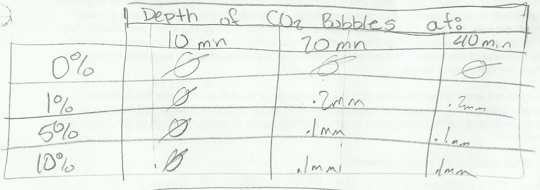
4. To test whether the concentration of sucrose affects the rate of alcoholic fermentation in yeast, you will measure the rate of CO2 productions for 4 different concentrations of sucrose. Complete the table to predict how much CO2 production you expect in each case.
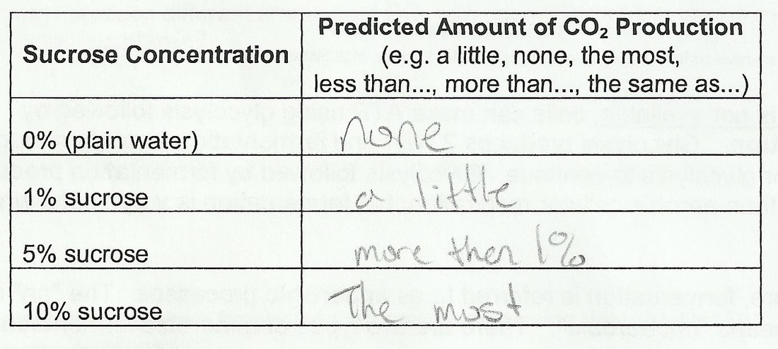
5. What will be the independent variable in your experiment?
The independent variable in the experiment is water.
What will be the dependent variable in your experiment?
The dependent variable in the experiment is sucrose.
The independent variable in the experiment is water.
What will be the dependent variable in your experiment?
The dependent variable in the experiment is sucrose.
6. What will be the control treatment in your experiment?
The control treatment in the experiment is plain water.
What is the purpose of this control treatment?
The purpose of the control treatment is to see how yeast affects the amount of sucrose.
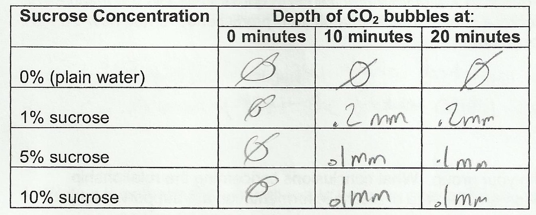
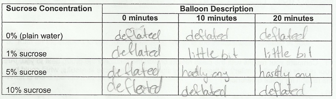
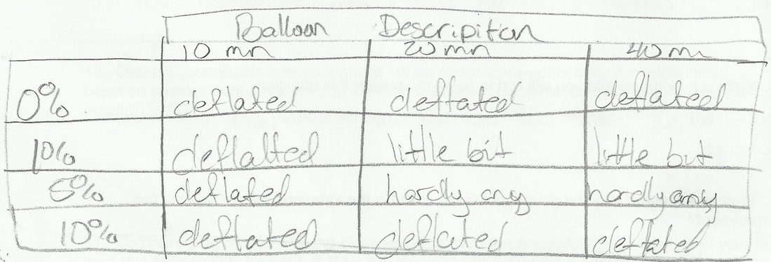
7. Record your observations in these data tables.
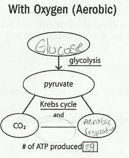
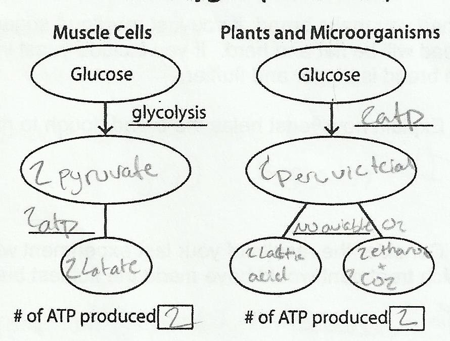
8. Use the information from page 1 to complete the figures below. Fill in the ovals with the appropriate molecule. On the blank lines write the name of the appropriate process. In the boxes at the bottom for the figure write how much ATP is made in each pathway.
|
|
9. What is the main advantage of aerobic respiration?
The main advantage of aerobic respiration is that it produces more ATP.
The main advantage of aerobic respiration is that it produces more ATP.
10. What is the main advantage of anaerobic fermentation?
The main advantage of anaerobic fermentation is that it requires no oxygen.
The main advantage of anaerobic fermentation is that it requires no oxygen.
11. Compare your results in question 7 with your predictions in question 4. Did the amounts of CO2 produced at different sucrose concentrations match your predictions? If not, how did the results differ from your expectations?
The 0% and 1% were just like we hypothesized, but the 5% and 10% produced less than what we thought.
The 0% and 1% were just like we hypothesized, but the 5% and 10% produced less than what we thought.
12. Discuss your results with your group. What conclusions concerning the relationship between sucrose concentration and the rate of alcoholic fermentation are supported by your results?
The less sucrose the faster the fermentation.
The less sucrose the faster the fermentation.
13. Compare your results with the class results or with the results of the group next to you. Are your results generally similar? If there are any significant differences in results, what could be the reason for these differences?
The group we talked with said they got the same results, that 1% did the best in the 20 minutes
The group we talked with said they got the same results, that 1% did the best in the 20 minutes
Experiment 2 - Yeast and Other Ingredients in Bread
1. Explain how yeast helps the bread dough to rise?
Yeast helps bread rise because it release CO2.
Yeast helps bread rise because it release CO2.
2. Consider the results of your last experiment with yeast and sucrose. If you add flour, which treatment would have made the fluffiest bread?
The fluffiest bread would come from the treatment with 10% because it release the most CO2 over time.
The fluffiest bread would come from the treatment with 10% because it release the most CO2 over time.
3. What question will you investigate?
Our question is how cinnamon will effect the fluffiness of bread
Our question is how cinnamon will effect the fluffiness of bread
4. Write a hypothesis that you will test to help you answer this question.
Our hypothesis is that the cinnamon will not effect the fluffiness of the bread.
Our hypothesis is that the cinnamon will not effect the fluffiness of the bread.
5. Plan an experiment to test your hypothesis. What is the independent variable in your experiment?
The independent variable is the water and the yeast.
The independent variable is the water and the yeast.
6. What is the control treatment in your experiment? (Hint: You are testing how sugar affects alcoholic fermentation so you will want to use 10% sucrose in each of your treatments).
The dependent variable is the cinnamon.
The dependent variable is the cinnamon.
7. Describe your procedures.
1) Fill test tubes with equal amounts of water.
2) Add 10% sucrose.
3) Add 0%, 1%, 5%, and 10% cinnamon into the test tubes
4) Add the yeast
5) Place a balloon on the test tubes immediately.
6) record data at 0, 10, 20, and 40 minutes.
1) Fill test tubes with equal amounts of water.
2) Add 10% sucrose.
3) Add 0%, 1%, 5%, and 10% cinnamon into the test tubes
4) Add the yeast
5) Place a balloon on the test tubes immediately.
6) record data at 0, 10, 20, and 40 minutes.
8. Create a data table or tables
9. Did the yeast produce different amounts of CO2 with your different treatments? Do the results match your hypothesis?
No the yeast didn't produce different amounts of CO2. Yes the results match my hypothesis.
No the yeast didn't produce different amounts of CO2. Yes the results match my hypothesis.
What do your results mean for people who make bread using the ingredient or temperatures you investigated?
My results show that use of cinnamon will not effect the fluffiness of bread.
My results show that use of cinnamon will not effect the fluffiness of bread.
G-AP BIOLOGY LAB 5 RESPIRATION: 09-26-14
Data and Analysis
Analysis
1. State a hypothesis that relates to temperature that is being tested by this lab exercise.
Analysis
1. State a hypothesis that relates to temperature that is being tested by this lab exercise.
2. State a hypothesis that relates to the state of seed germination that is being tested by this lab exercise.
3. Calculate the RATE of oxygen consumption for the germinating seeds in both cold and room temperature water. Rate can be calculated by determining the SLOPE of the line form your graph above.
4. In this lab exercise, what is the purpose of the ...
1. Beads
1. Beads
G-THE INTERNAL STRUCTURES OF CELLS:10-03-14
Cell Sizes
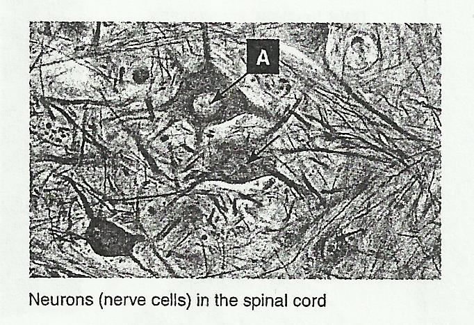
1. Using the measurement scales provided on each of the photographs above, determine the longest dimension (length or diameter) of the cell/animal/ virus in um and mm (choose the cell marked 'A' for epidermal cells)
(a) Amoeba: 100 um .1 mm
(b) Foraminiferan: 100 um .1mm
(c) Leprospira: 1 um .001 mm
(d) Epidermis: 50 um .5 mm
(e) Daphnia: 1000 um 1 mm
(f) Papillomavirus: .1 um .0001 mm
2. List these six organisms in order of sizes, from the smallest to the largest: Papillomavirus, Leptospira, Amoeba, Foraminiferan, Epidermis, Daphnia
3. Study the scale of your ruler and state which of these six organisms your would be able to see with your unaided eye: You could see a Daphnia because it is a millimeter.
4. Calculate the equivalent length in millimeters (mm) of the following measurements:
(a) 0.25 um: .00025 mm
(b) 450 um: .450 mm
(c) 200 nm: .0002 mm
Prokaryotic Cells
1. Describe three features distinguishing prokaryotic cells from eukaryotic cells:
(a) smaller
(b) lack nucleus and organelles
(c) simpler
2.
(a) Describe the function of flagella in bacteria: The flagella helps the bacteria move.
(b) Explain how fimbriae differ structurally and functionally from flagella: The fimbriae are around the whole cell, they are shorter, straighter, thinner, and used for attachment, not movement.
3.
(a) Describe the location and general composition of the bacterial cell wall: The location is behind the Glycocalyx, it is made of peptidoglycan, lipoproteins, and lipopolysaccharides,
(b) Describe how the glycocalyx differs from the cell wall: The glycocalyx is gelatinous, and it attaches to a substrate.
4.
(a) Describe the main method by which bacteria reproduce: Bacteria divide by binary fission, this is where DNA is copied and the cell splits.
(b) Explain how conjugation differs from this usual method: Conjugation involves two bacteria with one way genetic transfer.
5. Briefly describe how the artificial manipulation of plasmids has been used for technological applications: Artificial Manipulation of plasmids are used for cloning.
Animal Cells
1. Explain what is meant by a generalized cell: A general cell is a cell that shows all the organelles of a cell for it to properly function.
2. Discuss how the shape and size of a specialized cell, as well as the number and types of organelles it has, are related to its functional role. Use examples to illustrate your answer: The size depends on the number of organelles that the cell needs to function, the shape determines where it is in the body and what is is doing. Skin cells hook to each other to make the layers of skin.
3. The two photomicrographs (below) show several types of animal cells. Identify the features indicated by the letters A-C:
A: Nucleus
B: White blood cell
C: White blood cell
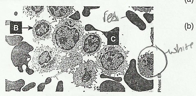
4. White blood cells are mobile, phagocytic cells, whereas red blood cells are smaller than white blood cells and, in humans, lack a nucleus.
(a) In the photomicrograph (below), circle a white blood cell and a red blood cell:
(a) In the photomicrograph (below), circle a white blood cell and a red blood cell:

(b) With respect to the features that you can see, explain how you made your decision: It is a white blood cell because you can see organelles, and the other is a red blood cell because you can't see organelles.
5. Name and described one structure or organelle present in generalized animal cells but absent from plant cells: Centrioles are used by animals cells in nuclear division.
5. Name and described one structure or organelle present in generalized animal cells but absent from plant cells: Centrioles are used by animals cells in nuclear division.
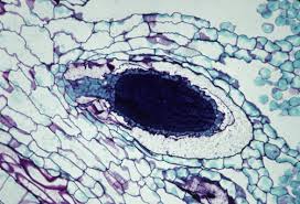
Plant Cells
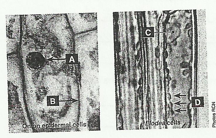
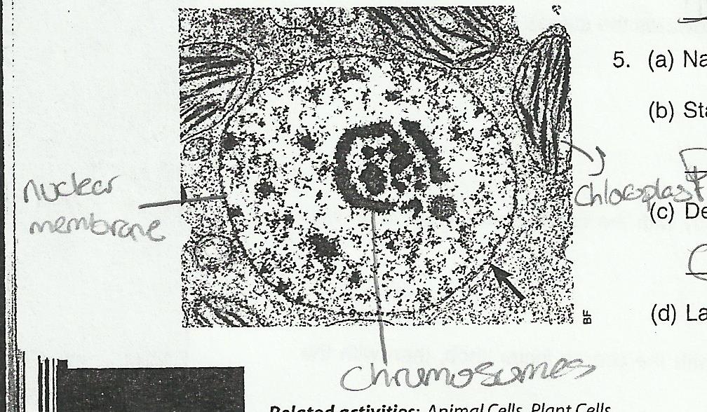
1. The photographs (below) show plant cells as seen by a light microscope. Identify the basic features labelled A-D:
A: Starch Granule
B: Cell Wall
C: Nucleus
D: Chloroplast
1. The photographs (below) show plant cells as seen by a light microscope. Identify the basic features labelled A-D:
A: Starch Granule
B: Cell Wall
C: Nucleus
D: Chloroplast
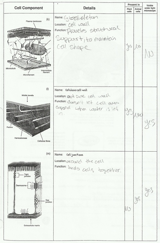
2. Describe three structures/organelles present in generalized plant cells but absent from animal cells:
(a) Chloroplast is used in photosynthesis.
(b) Cell Wall is used for shaping the cell and volume control.
(c) Starch Granule is used for storage.
(a) Chloroplast is used in photosynthesis.
(b) Cell Wall is used for shaping the cell and volume control.
(c) Starch Granule is used for storage.
The Cell's Cytoskeleton
1. Describe the role that all components of the cytoskeleton have in common: All the components of the cytoskeleton help in maintaining the cell shape.
2. Explain the importance of the cytoskeleton being a dynamic structure: The cytoskeleton changes the shape, move material, and moves the cell.
3. Explain how the presence of a cytoskeleton could aid in directing the movement of materials within a cell: The cytoskeleton is what moves material through the cell by moving the shape of the cell itself.
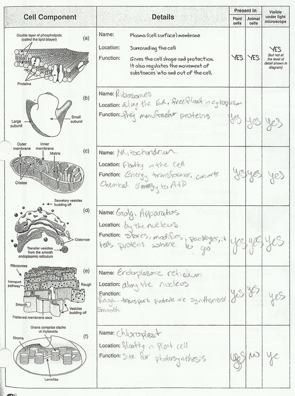
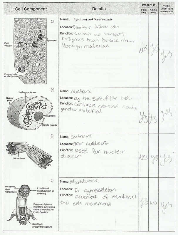
Cell Structures and Organelles
Cell Processes
1. For each of the processes listed below, identify the organelle(s) or structure(s) associated with that process.
(a) Secretion: Golgi Apparatus
(b) Respiration: Cytoplasm and Mitochondria
(c) Pinocytosis: Plasma Membrane
(d) Phagocytosis: Plasma Membrane
(e) Protein Synthesis: Ribosome in the Cytoplasm and on the Endoplasmic Reticulum
(f) Photosynthesis: Chloroplast
(g) Cell Division: Centrioles
(h) Autolysis: Lysosomes
(i) Transport in/out of cell: Plasma Membrane
2. Explain what is meant by metabolism and describe an example of a metabolic process: Metabolism is the sum of all the processes of a cell.
Golgi and the Endoplasmic Reticulum
1. Using examples, explain what is meant by a macromolecule: A macromolecule is a organic polymer made up of repeating units of smaller molecules
2. Why are polypeptides requiring transport synthesized by the membrane-bound (rather than free) ribosomes?
Polypeptides require transport synthesis by membrane-bound ribosomes because the polypeptides goes into the Endoplasmic Reticulum that the Ribosomes are attached to.
3. Why are most proteins destined for secretion from the cell glycoproteins?
Most of the proteins destined for secretion are glycoproteins because carbohydrates are destined to be secreted and glycoproteins are just proteins with carbohydrates attached to them.
4. Briefly describe the roles of the following organelles in the production of macromolecules:
(a) Rough ER: The Rough ER assembles proteins for secretion.
(b) Smooth ER: The Smooth ER synthesis fats, phospholipids, steroid hormones, and other lipids.
(c) Golgi Apparatus: The Golgi Apparatus receives transport vesicles containing export materials, and produces vesicles.
(d) Transport Vesicles: The Transport Vesicle takes secretion materials to the Golgi Apparatus to by taken outside the cell.
Identifying Structures in an Animal Cell
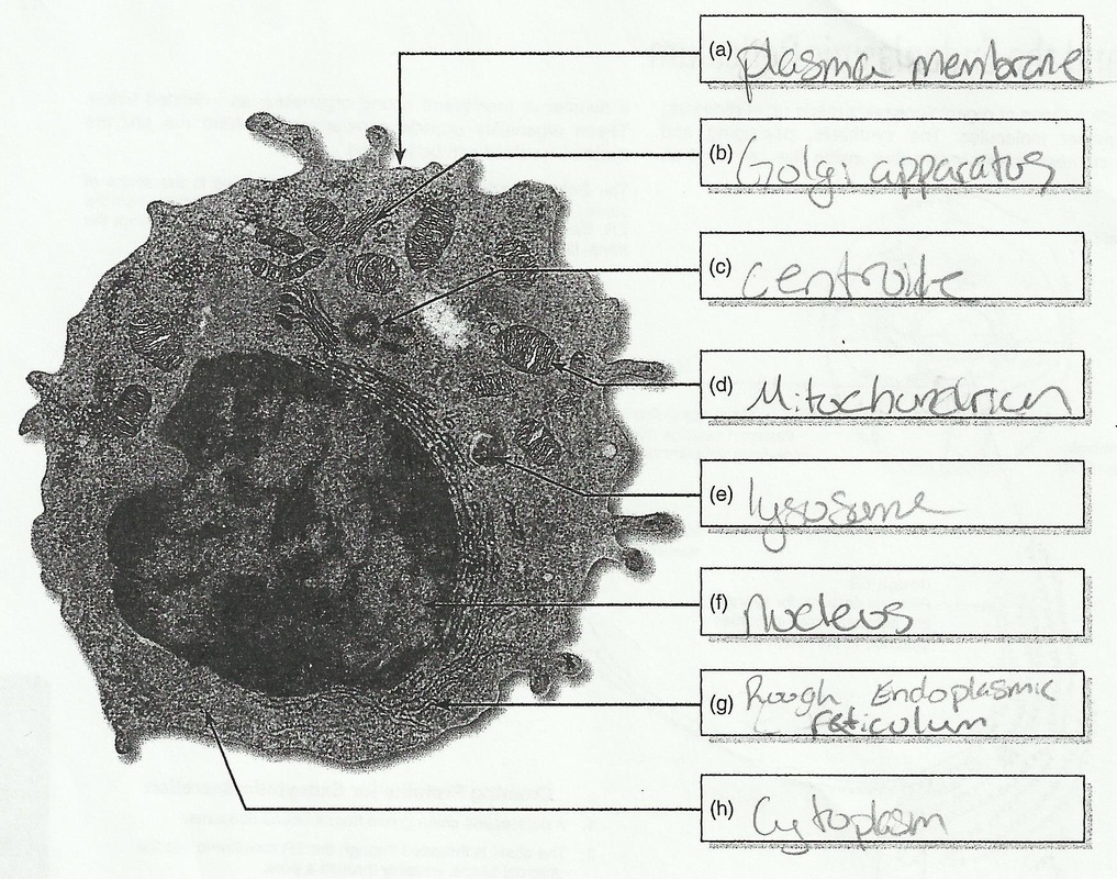
1. Identify and label the structures in the cell above using the following list of terms: cytoplasm, plasma membrane, rough endoplasmic reticulum, mitochondrion, nucleus, centriole, Golgi apparatus, lysosome
2. Which of the organelles in the EM above are shown in both transverse and longitudinal section?
The Nucleus, Plasma Membrane, and the Cytoplasm are shown in both transverse and longitudinal section.
3. Why do plants lack the mobile phagocytic cells typical of animal cells?
Plant cells lack the mobile phagocytic cells typical of animal cells because they don't have to move anywhere in the plant like animal cell do.
4. The animal cell pictured above is a lymphocyte. Described the features that suggest to you that:
(a) It has a role in producing and secreting proteins: You can it has a role in producing and secreting proteins because it has rough endoplasmic reticulum, ribosomes, and golgi apparatus, all of these in protein secretion.
(b) It is metabolically very active: You can see that it is metabolically very active because it has many organelles and the sum of the processes will be greater than that of other cells with less organelles.
5. What features of the lymophocyte cell above identify it as eukaryotic?
The lymophocyte cell has organelles and a visible nucleus, prokaryotic cells have neither of these.
Identifying Structures in a Plant Cell
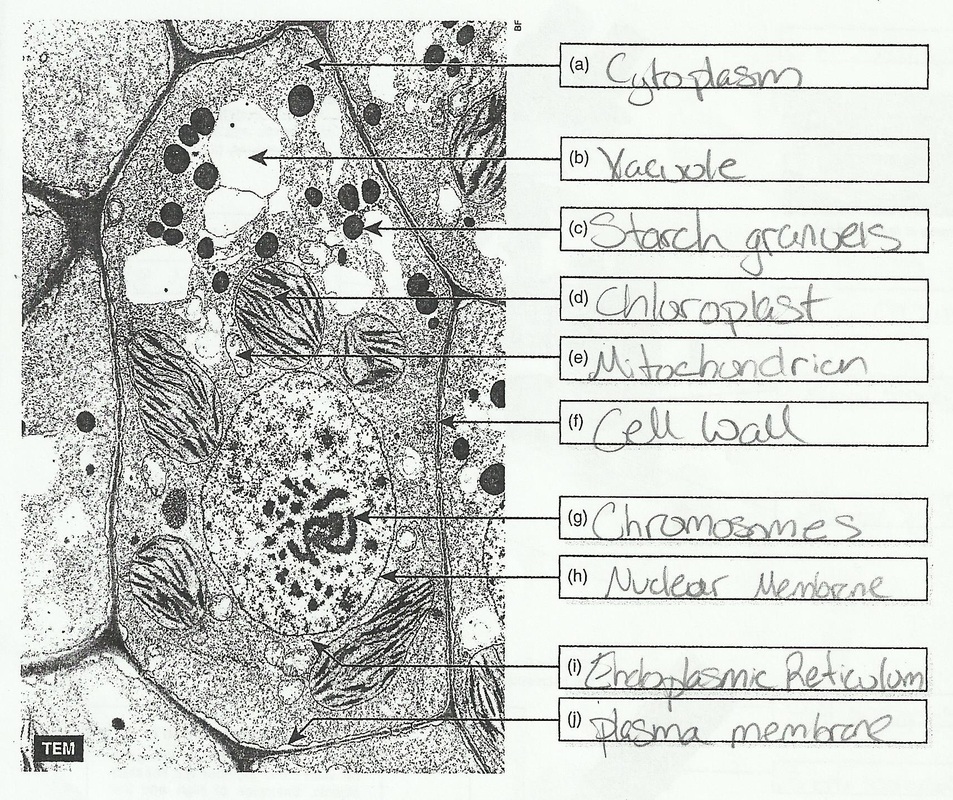
1. Study the diagrams on the other pages in this chapter to familiarize yourself with the structures found in eukaryotic cells. Identify and label the ten structures in the cell above using the following list of terms: nuclear membrane, cytoplasm, endoplasmic reticulum, mitochondrion, starch granule, chromosome, vacuole, plasma membrane, cell wall, chloroplast.
2. State how many cells, or parts of cells, are visible in the electron microscope above: There are nine cells or parts of cells that are visible in the electron microscope above
3. Describe the features that identify this cell as a plant cell: Chloroplast and Cell Walls only belong to plant cells.
4.
(a) Explain where cytoplasm is found in the cell: Cytoplasm is found all through the cell.
(b) Describe what cytoplasm is made up of: Cytoplasm is made up of a watery substance containing dissolved substances, enzymes, and the cell organelles and structures.
5. Describe two structures, pictured in the cell above, that are associated with storage:
(a) Vacuole stores water for equilibrium.
(b) Chloroplast stores the chlorophyll for photosynthesis.
Optical Microscopes
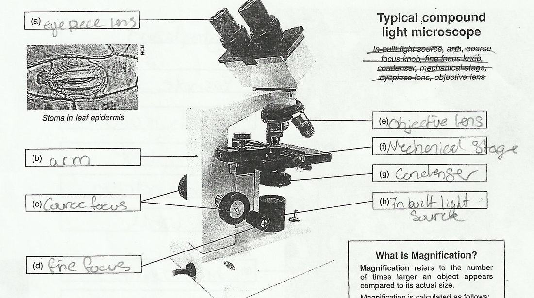
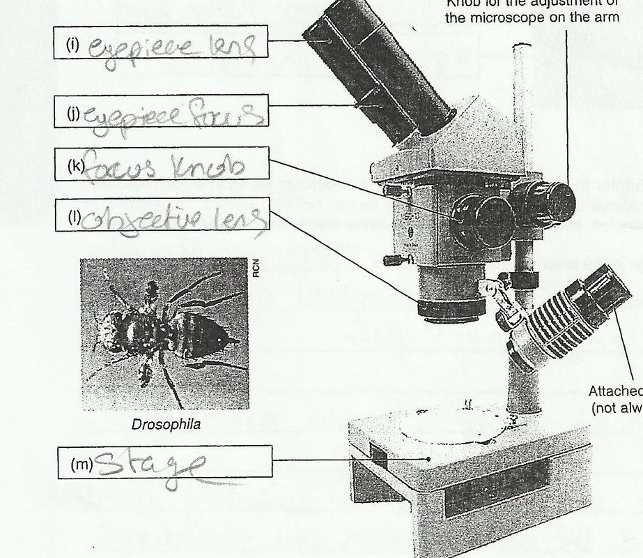
1. Label the two photographs, the compound light microscope (a) to (h) and the dissecting microscope (i) to (m). Use words from the lists supplied for each image.
Typical Compound Light Microscope: In-built light source, Arm, Course focus-knob, Fine focus-knob, Condenser, Mechanical stage, Eyepiece lens, Objective lens
Dissecting Microscope: Focus knob, Stage, Eyepiece lens, Objective lens, Eyepiece focus
2. Determine the magnification of a microscope using
(a) 15 X eyepiece and 40 X objective lens: 55 Magnification
(b) 10 X eyepiece and 60 X objective lens: 70 Magnification
3. Describe the main difference between a compound light microscope and a dissecting microscope: A dissecting microscope is used for low magnification while a compound light microscope is used for higher magnification.
4. What type of microscope would be used to:
(a) Count stream invertebrates in a sample: Dissecting
(b) Observe cells in Mitosis: Compound Light
5.
(a) Distinguish between magnification and resolution (resolving power): Magnification is the number of times larger of an image, resolution makes an image more or less clear.
(b) Explain the benefits of higher resolution: With higher resolution you can see an image more clearly than with a lower resolution.
6. Below is a list of ten key steps taken to set up a microscope and optimally view a sample. The steps have been mixed up. Put them in their correct order by numbering each step.
9) Focus and center the specimen using the high objective lens. Adjust focus using the fine focus knob only.
5) Adjust the illumination to an appropriate level by adjusting the iris diaphragm and the condenser. The light should appear on the slide directly below the objective lens, and give an even amount of illumination.
1) Rotate the objective lenses until the shortest lens is in place (pointing down towards the stage). This is the lowest power objective lens.
3) Place the slide on the microscope stage. Secure with the stage clips.
10) Fine tune the illumination so you can view maximum detail on your sample.
8) Focus and center the specimen using the medium objective lens. Focus firstly with the course focus knob, then with the fine focus knob (if needed).
4) Turn on the light source.
7) Focus and center the specimen using the low objective lens. Focus with the course focus knob, then with the fine focus knob.
6) Focus the eyepieces to adjust your view.
2) Adjust the distance between the eyepieces sot hat they are comfortable for your eyes.
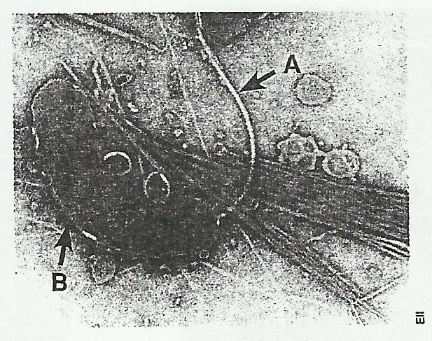
Interpreting Electron Micrographs
|
1.
(a) State which kind of cell this is: Prokaryotic (b) Identify the structure labelled A: Flagellum (c) Describe the function on this structure: Moves the cell (d) Identify the structure labelled B: Glycocalyx |
|
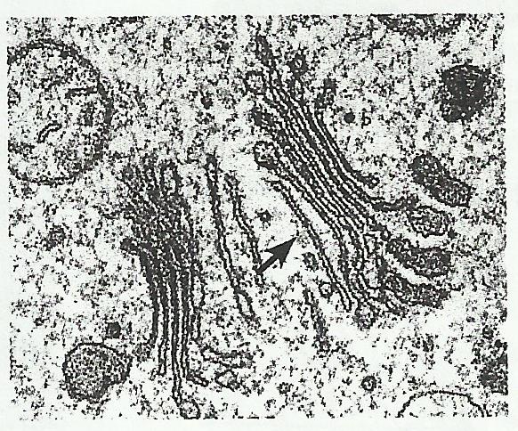
2.
(a) Name this organelle (arrowed): Golgi Apparatus (b) State which kind of cell(s) this organelle would be found in: plants and animals (c) Describe the function of this organelle: Stores, modifies, and packages proteins. |
|
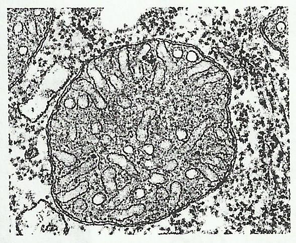
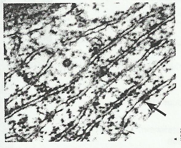
3.
(a) Name the large, circular organelle: Mitochondrion (b) State which kind of cell(s) this organelle would be found in: plants and animals (c) Describe the function of this organelle: transforms energy (d) Label two regions that can be seen inside the organelle |
|
4.
(a) Name and label the ribbon-like organelle in this photograph (arrowed): Cell Wall (b) State which kind of cell(s) this organelle is found in: plants and animals (c) Describe the function of these organelles: to regulate the flow of material in and out of the cell (d) Describe the dark 'blobs' attached to the organelle you have labelled: Starch Granules |
|
5.
(a) Name the large circular structure (arrowed): Nucleus (b) State which kinds of cell(s) this organelle is found in: plants and animals (c) Describe the function of this organelle: Controls the cell, hold genetic material (DNA) (d) Label three features relating to this structure in the photograph. |
Microscopy Techniques
1. Why are many microscopes samples wet mounted prior to viewing under a microscope?
A wet mount improves the appearance and enhances detail.
2. What is the main purpose of using stain?
Stains help highlight specific structures.
3. What is the difference between a viral and non-viable stain?
Vital stains are used on live specimens while non-viable stain are used on non-living specimens.
4. Identify a stain that would be appropriate for improving identification of the following:
(a) Fungal hyphae: Trypan Blue
(b) Starch in potatoes: Iodine Solution
(c) Cell Wall of bacteria: Crystal Violet
(d) Lignin in a plant root section: Aniline Sulfate
(e) Nuclei in cheek cells: Methylene Blue
Calculating Linear Magnification
1. The bright field microscopy image on the left is of onion epidermal cells. The measured length of onion cell in the center of the photograph is 52,000 um (52 mm). The image had been magnified 140 X. Calculate the actual size of the cell:
x= 52mm divided by 140
x= .37mm
2. The image of the flea (left) has been captured using light microscopy.
(a) Calculate the magnification using the scale line on the image:
x= 2mm divided by .5mm
x= 4mm
(b) The body length of the flea is indicated by a line. Measure along the line and calculate the actual length of the flea:
2mm divided by 4mm = .5mm
3. The image size of the E.coli cell (left) is 43mm, and its actual length is 2 um. Using this information, calculate the magnification of the image:
x= 43,000um divided by 2um
x= 21,500 um
Making Biological Drawings
1. Identify and describe eight unacceptable features of the student's biological diagram above:
(a) Labeling lines cross each other
(b) Lines are not drawn with ruler
(c) The drawing is in the corner not the center
(d) The drawing is small
(e) It is not of the whole drawing
(f) Proportions not accurate
(g) They didn't put a scale
(h) Simple narrow lines are not used
2. In the remaining space next to the 'poor example' (above) or on a blank sheet of paper, attempt your own version of a biological drawing for the same material, based on the photograph above. Make a point of correcting all of the errors that you have identified in the sample student's attempt.
3. Why is an accurate biological drawing more valuable to a scientific investigation than an 'artistic' one?
A biological drawing in more valuable to scientific investigation than an 'artistic' one because the 'artistic' one is up to interpretation instead of what is really there.
SHOW ME YOUR TATTOO: 10-12-14
Summer is the best time to get a tattoo; you get to show it off when out in the sun. Well you might want to rethink going out for fun in the sun with that new tattoo of yours. All tattoos will fade, some faster than others, but the sun will fade them faster than just with time alone. Tattoo ink particles are relatively large but when exposed to sun light the particles shrink allowing your skin to wash them out causing fading. Brighter ink or cheaper get broken down faster than darker or better quality inks. Tanning beds also dulls the vibrancy; some say it is worse because the rays are concentrated. To keep that tattoo looking the best when out in the sun put on a good sunscreen and wear long sleeve heavier material shirts. UV rays can go through some materials and take the vibrancy from the tattoo: if you hold a shirt up to the sun and can see light through it the sun can reach the tattoo. You need to also moisturize the skin under and around the tattoo. If the tattoo is new follow the care directions from the artist and stay out of the sun until the tattoo is completely healed.
Blog. (2013, May 30). Retrieved October 6, 2014, from http://www.revitaink.com/tattoo/sun-and-tattoos-dont-mix/
Guide Line Tattoos - Care & Feeding of a New Tattoo. (n.d.). Retrieved October 6, 2014, from http://www.guidelinetattoos.com/care.html
Snyder, M. (2012, July 22). Tattoos and sun exposure: A losing combination. Retrieved September 26, 2014, from http://onmilwaukee.com/living/articles/tattoosandsun.html
The Sun's Damaging UV Rays and Dry Skin Fade the Bright Colors of Tattoos. (2008, January 1). Retrieved October 11, 2014, from http://www.saveyourtattoo.com/whytattoosfade.html
Unit 3: Akanthos
Floating Leaf Disk Photosynthesis Lab: 10-23-14
Purpose: The purpose is to see how different light intensity effect photosynthesis.
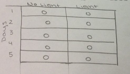
Predictions:
How will the different light conditions affect the rate of floating disks?
The disks in the light will have the most floating disks, the disks in ambient light will have some, and disks in the dark will have none.
How will the different light conditions affect the rate of floating disks?
The disks in the light will have the most floating disks, the disks in ambient light will have some, and disks in the dark will have none.
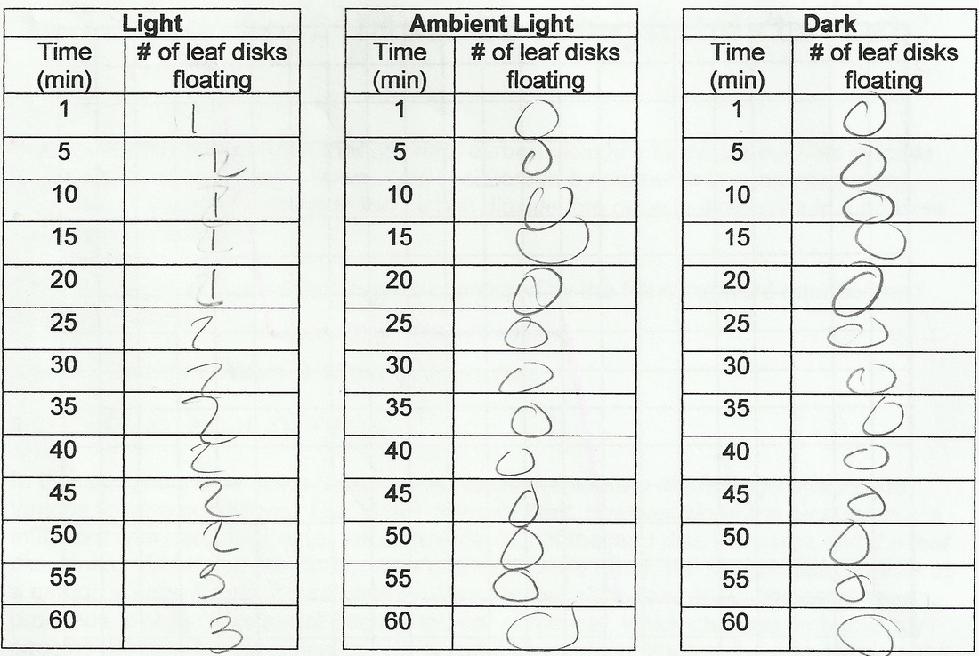
Qualitative Observations: The disks in the light had some disks floating and had soap bubbles. The disks in both ambient light and in the dark had none floating and no soap bubbles.
1. Graph your results for all three trials in the graph paper below. Label the graph, both axes and provide a legend to distinguish each trial.
2. What was the role of the sodium bicarbonate in this experiment?
3. Which trial resulted in all the leaf disks floating the fastest? Explain.
4. Explain the process that caused the leaf disks to rise.
5. If the leaf disks were boiled, what kind of results would you expect?
6. How does light intensity affect the rate of photosynthesis?
7. The same experiment was conducted where 10 leaf disks were placed in a sodium bicarbonate solution and placed in the light. Every minute, the number of floating disks were counted and recorded. After 14 minutes, the leaf disks were floating.
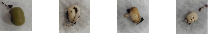
POLLEN TUBE GROWTH: 10-23-14
Before
After
GERMINATION INHIBITORS: 10-24-14
Analysis Questions:
1. What are the control(s) in your experiment?
The control was the seeds in just the water.
Why did you wash the seeds?
We washed the seeds to make sure the control wasn't effected by the tomato juice.
The control was the seeds in just the water.
Why did you wash the seeds?
We washed the seeds to make sure the control wasn't effected by the tomato juice.
2. Explain how you determined whether or not a seed has germinated.
You can tell if a seed has germinated or not by if it grows over a period of time.
You can tell if a seed has germinated or not by if it grows over a period of time.
3. What conclusions can you draw from the results: do tomatoes contain germination inhibitors?
Yes, tomatoes do contain germination inhibitors.
Do these inhibitors work for other types of tomatoes?
Yes, these inhibitors work for other tomatoes as well.
Yes, tomatoes do contain germination inhibitors.
Do these inhibitors work for other types of tomatoes?
Yes, these inhibitors work for other tomatoes as well.
4. In addition to tomatoes, what other fruits or vegetables probably contain germination inhibitors?
Any types of fruits or vegetables with seeds in them would contain the seed germination inhibitors.
Any types of fruits or vegetables with seeds in them would contain the seed germination inhibitors.
5. Discuss some practical applications of germination inhibitors.
A practical use of a germination inhibitor would be to make sure the seeds don't grow in the already grown plants.
A practical use of a germination inhibitor would be to make sure the seeds don't grow in the already grown plants.
SEED GERMINATION EXPERIMENTS: 10-24-14
Experiment one:
1. The question:
How does acid affect the germination of a seed?
How does acid affect the germination of a seed?
2. The hypothesis:
The more acid added the less the seed will germinate.
The more acid added the less the seed will germinate.
3. The experimental design:
Materials:
4 test tubes
4 not germinated seeds
Hydrochloric(HCl) acid
Gradulated cylinder
Soil
Water
Procedure:
1) Label test tubes 1,2,3, and 4.
2) Put 2 inches of soil in each test tube then put one seed in each, add one more inch of soil on top.
3) Put test tubes in test tube holder.
4) In test tube one add nothing.
5) In test tube two add 10 mL of water.
6) In test tube three add 5 mL of water and 5 mL of HCl.
7) In test tube four add 10 mL of HCl.
8) Create a data table for a week long experiment.
9) Repeat steps 4-7 daily and record observations.
4. Data:
5. Conclusions:
The plant with 10 mL of water grew the best like predicted but the plant with 10 mL of acid also germinated and grew instead of killing the seed like we originally thought.
The plant with 10 mL of water grew the best like predicted but the plant with 10 mL of acid also germinated and grew instead of killing the seed like we originally thought.
Non-germinated, 10mL of water, Half water half acid, 10 mL of acid
Experiment two:
1. The question:
How is the germination of a seed affected by flooding?
How is the germination of a seed affected by flooding?
2. The hypothesis:
The seed will not grow with more flooding.
The seed will not grow with more flooding.
3. The experimental design:
The experimental design:
Materials:
4 test tubes
4 not germinated seeds
Gradulated cylinder
Soil
Water
Procedure:
1) Label test tubes 1,2,3, and 4.
2) Put 2 inches of soil in each test tube then put one seed in each, add one more inch of soil on top.
3) Put test tubes in test tube holder.
4) In test tube one add nothing.
5) In test tube two add 10 mL of water.
6) In test tube three add 20 mL of water.
7) In test tube four add 30 mL of water.
8) Create a data table for a week long experiment.
9) Repeat steps 4-7 daily and record observations.
4. Data:
5. Conclusions:
The 10mL of water grew the best, the 20mL and 30mL grew fairly well, but not as much since they had too much water constantly. This flooding caused the seeds to have trouble in their growing processes.
The 10mL of water grew the best, the 20mL and 30mL grew fairly well, but not as much since they had too much water constantly. This flooding caused the seeds to have trouble in their growing processes.
WHERE DO PLANTS GET THEIR FOOD?: 10-24-14
A.
Hypothesis: Soil on its own will not provide the need nutrients for the plant to grow.
Hypothesis: Soil on its own will not provide the need nutrients for the plant to grow.
B.
1) Take two germinating seeds.
2) Plant them in separate pots.
3) Place one under light and the other in complete darkness.
4) Water once a day with 40 mL of water.
5) Repeat step 4 for one week.
1) Take two germinating seeds.
2) Plant them in separate pots.
3) Place one under light and the other in complete darkness.
4) Water once a day with 40 mL of water.
5) Repeat step 4 for one week.
D. The plants did not consume the soil because over the one week period there was on growth of either plant.
PHOTOSYSTEMS AND CHEMIOSMOSIS - THE MECHANISM OF ATP SYNTHESIS IN CHLOROPLASTS
1. Explain or describe in words, the path electrons take through the thylakoid.
Electrons leave photosystem II when they are excited by light. They enter the eletron transport chain and are passed through a series of proteins, PQ, b6-f, Pc. This process pumps protons across the membrane against their concentration gradient. Light also hits photosystem I where electrons are passed to the protein ferredoxin. As the electron is passed down that chain NADPH is produced.
Electrons leave photosystem II when they are excited by light. They enter the eletron transport chain and are passed through a series of proteins, PQ, b6-f, Pc. This process pumps protons across the membrane against their concentration gradient. Light also hits photosystem I where electrons are passed to the protein ferredoxin. As the electron is passed down that chain NADPH is produced.
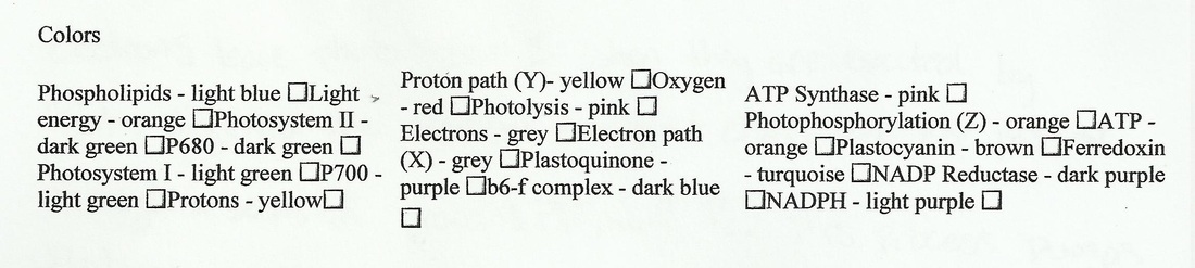
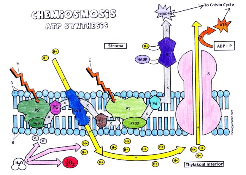
2. Explain the role of each of the following:
- Plastocyanin - protein that is the final electron receptor in the electron transport chain
- Plastoquinone - the first electron acceptor in the ETC
- Plastocyanin - protein that is the final electron receptor in the electron transport chain
- Plastoquinone - the first electron acceptor in the ETC
